Cosmetic Dentistry
White Spots on Teeth: What Causes Them and Can They Be Fixed?
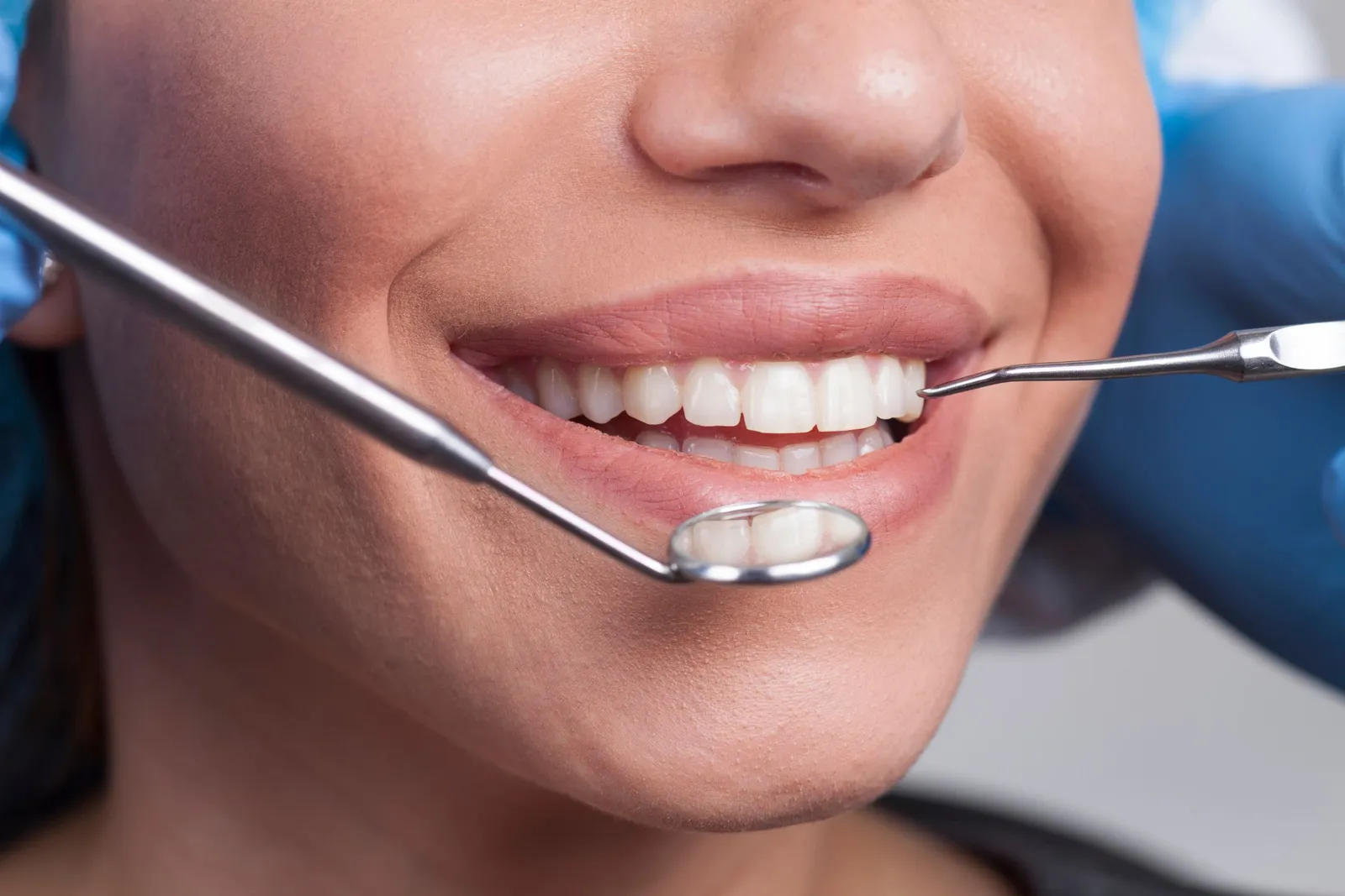
A white spot is not a stain sitting on your tooth. It is a patch of enamel that is more porous than the enamel around it, and it looks white for the same reason snow does: light scatters inside it instead of passing through. That is why scrubbing harder or switching to a whitening toothpaste never makes a dent.
What does work depends on why the spot is there. Three causes account for most of them, each responds to different treatment, and sorting out which one you have is the first job of a cosmetic dental exam.
What causes white spots on teeth, and can they be fixed?
Short answer: Most white spots come from one of three things: mineral loss under plaque (very common after braces), too much fluoride swallowed while the teeth were forming, or enamel that developed imperfectly in childhood. Nearly all can be improved. Fresh post-braces spots often fade with fluoride and time; stubborn ones usually respond to microabrasion, resin infiltration or bonding, frequently with no drilling.
The main causes, and how to tell them apart
Demineralization: the chalky marks left after braces
Plaque that sits against enamel for weeks produces acid, and that acid pulls calcium and phosphate out from just beneath the surface. The outer skin of the enamel stays intact while the layer under it turns porous. Dentists call the result a white spot lesion.
Brackets make this easy. They create ledges a toothbrush skips, so the classic pattern is a chalky frame around where each bracket sat, or a dull white band hugging the gumline. It can happen faster than people expect — a spot can form within about a month of poor cleaning around a bracket.
You do not need braces to get them. The same lesions show up along the gumline of anyone who leaves plaque there, especially with a dry mouth or a habit of sipping soda or sports drinks through the day.
Fluorosis: faint, symmetrical streaks
Fluorosis happens when a child takes in more fluoride than needed while the permanent teeth are still forming under the gums, a window the CDC puts at roughly the first eight years of life. Swallowed toothpaste is the usual culprit. Once a tooth has come in, it cannot develop fluorosis.
The great majority of cases in the United States are very mild or mild: lacy white lines or small flecks, typically mirrored on matching teeth on the left and right. It is not decay and the teeth are not weaker, a point the ADA's MouthHealthy guide to fluorosis makes plainly. The rarer moderate and severe forms bring brown mottling and pitted enamel.
Enamel hypoplasia and hypomineralization: the spot that was always there
Sometimes enamel forms thin or under-mineralized because something interrupted it. A high fever or serious illness in early childhood can do it. So can a knock to a baby tooth, or an infection in one, while the adult tooth was developing right above it. Often nobody can point to a reason.
The giveaway is history: the mark was there the day the tooth came in. It usually affects one tooth or a few, has a sharp border, and ranges from creamy white to yellow-brown, sometimes with a pit you can feel. These spots tend to run deeper through the enamel than the other two types. That matters a great deal once you start talking about treatment.
The spots that vanish by mid-morning
If your front teeth look blotchy when you wake up and normal by the time you have had breakfast, that is dehydration. Sleeping with your mouth open dries the enamel, and dry enamel shows every faint irregularity. Saliva rewets it and the blotches fade. Harmless — though constant mouth breathing is worth mentioning at your next visit.
White spots at a glance: cause, appearance, what tends to work
| Cause | What it looks like | Is it decay? | What tends to work |
|---|---|---|---|
| Demineralization (often after braces) | Chalky, matte patches around old bracket positions or along the gumline | Yes — the earliest stage, and usually stoppable | Fluoride and time for fresh spots; resin infiltration or microabrasion for what remains |
| Mild fluorosis | Faint lacy lines or flecks, mirrored on matching teeth | No | Often nothing at all; whitening to blend, microabrasion or resin infiltration |
| Moderate to severe fluorosis | Dense white mottling, brown staining, sometimes pitting | No | Microabrasion with whitening; bonding or veneers where enamel is pitted |
| Enamel hypoplasia or hypomineralization | One or a few sharply bordered spots, cream to yellow-brown, present since the tooth erupted | No, but the weaker enamel decays more easily | Bonding most often; infiltration for shallow spots; veneers for widespread defects |
| Dehydration | Blotches on waking or right after whitening that fade within hours | No | Nothing |
Are white spots cavities?
Some of them are the first stage of one. A demineralization spot is tooth decay before there is a hole. The surface is still intact, no filling is needed yet, and the process can be halted and partly reversed. Fluorosis and developmental spots are not decay at all.
For a demineralized spot, the question that matters is whether it is active or arrested. An active lesion looks chalky and dull even when wet, feels faintly rough, and sits where plaque collects; the gum beside it is often puffy or bleeds when you brush. An arrested lesion is smooth, hard and glossy. It is a scar. It may bother you in photos, but it is not going anywhere.
A white spot needs to be treated as decay, not as a cosmetic problem, when:
- it is turning tan or brown and feels soft or sticky
- the surface has broken — a pit, or an edge that catches floss or a fingernail
- the tooth zings with sweets or cold drinks
- the spot is growing, or new ones keep appearing
- there is visible plaque on it and the gum next to it bleeds
A spot that has broken through needs a tooth-colored filling. An active one with an intact surface needs fluoride, better cleaning in that area, and a recheck. Cosmetic work placed over an active lesion is decoration on top of a problem, so the cause gets fixed first.
What happens if you wait? With an arrested scar, nothing. You can leave it for years and decide later. An active lesion is different: left under plaque it commonly progresses to a true cavity over months to a few years, and faster with a dry mouth or frequent sugar.
One distinction worth making: all of this is about spots on teeth. A white patch on the gums, tongue or inside of the cheek that does not wipe off and has not cleared in two weeks is a separate matter and should be looked at during an oral cancer screening.
Does whitening fix white spots or make them more obvious?
Both, depending on the week you look.
Bleaching gel lightens the whole tooth, spots included. It also dehydrates enamel temporarily, and dehydrated porous enamel looks even whiter. So for the first few days after whitening — occasionally as long as two weeks — the spots stand out more than they did before. This is the point where people panic. Then the teeth rehydrate and the contrast settles back.
Where it settles depends on the spot. Faint fluorosis lines on teeth that were fairly yellow often blend in nicely, because the background shade has moved toward the color of the spot. Dense, opaque patches stay visible. They were whiter than the rest of the tooth before, and they still are.
If you plan to have both professional whitening and a spot treatment, the whitening goes first. Resin and bonding material do not bleach, so they have to be matched to your final shade. Dentists commonly wait around two weeks after whitening before bonding anything, both for the color to stabilize and because freshly bleached enamel does not hold a bond as reliably. Some zingy sensitivity in the meantime is normal; we cover that in what to expect after whitening.
Treatment options a dentist may discuss
These run from least to most invasive. A good plan starts at the top and only moves down the list if it has to.
- Remineralization. Fluoride varnish, prescription-strength fluoride toothpaste or a calcium-phosphate paste, combined with fixing whatever let plaque sit there. This suits post-braces spots in the first few months, when saliva is already repairing them unaided; many shrink noticeably in the three to six months after brackets come off. The honest limit: it hardens a lesion reliably and erases the whiteness less reliably. Spots that are years old rarely disappear with fluoride alone.
- Microabrasion. A mild acid-and-abrasive paste is polished across the enamel to remove a very thin outer layer, a fraction of a millimeter. It works on shallow defects and on fluorosis, and is often paired with whitening. It does little for spots that go deep.
- Resin infiltration. The surface is conditioned with an etching gel and dried, then a very thin clear resin is drawn into the pores and hardened with a curing light. Filling those pores changes how light travels through the spot, so it takes on the look of the enamel around it. There is no drilling and usually no numbing. Results tend to be best on post-braces lesions and mild fluorosis, and less predictable on deep developmental spots, which may only partly mask. Not every office provides it, so ask.
- Composite bonding. For deeper spots or pitted enamel, the defective layer is thinned and replaced with tooth-colored resin in a single visit. Dental bonding hides what infiltration cannot, with trade-offs: composite picks up stain, wears, and typically needs polishing or replacing after some years. Matching one front tooth to its neighbor also takes real skill.
- Veneers. Porcelain veneers make sense for severe fluorosis or hypoplasia spread across several front teeth, or when shape needs correcting too. Enamel is removed permanently and you are committing to replacements for life. For one small spot it is almost always more dentistry than the problem deserves. The reasoning mirrors what we lay out in veneers versus bonding for a chipped front tooth.
What shifts the cost
Four things, mostly: how many teeth are involved, how deep the spots go (which decides whether a surface treatment is enough or bonding is needed), whether whitening comes first, and how many visits it takes. Dental plans generally class spot-masking as cosmetic and do not pay for it, while treating active decay with fluoride or a filling usually falls under preventive or restorative benefits. Our guide to insurance coverage for cosmetic work explains where that line tends to sit.
Questions worth asking before you agree to anything
- Is this spot active or arrested?
- How deep does it go, and how can you tell?
- Should I whiten first, and how long do we wait afterward?
- How much enamel does this option remove? Is it reversible?
- What will it look like in five years, and what upkeep does it need?
- What happens if we do nothing?
How to prevent white spots during braces or aligners
With brackets on, the strip of enamel between the bracket and the gumline is where nearly all of these form. Aim there.
- Brush after every meal, angling the bristles above the bracket and then below it, not just across the front.
- Use fluoride toothpaste and spit without rinsing, so the fluoride stays on the teeth. Add a daily fluoride rinse if your dentist or orthodontist recommends one.
- Clean under the wire with an interdental brush. A water flosser helps with what is left.
- Keep sweet and acidic drinks to mealtimes. How often your teeth are bathed in sugar matters more than how much.
- Stay on schedule with professional cleanings throughout orthodontic treatment. It is the visit people skip, and the one where early spots get caught and fluoride varnish goes on.
- Check the mirror weekly. Puffy red gums and a dull white line along the gum margin are the early warning. Cleaned up at that stage, the damage often reverses.
Clear aligners carry less risk because the trays come out and you can brush normally. Less is not zero. The classic mistake is drinking anything other than water with trays in: sweet or acidic liquid seeps under the plastic and sits against the enamel with no saliva to wash it away. Brush before the trays go back in after eating, and clean the trays themselves daily. If you are still choosing between systems, our Invisalign page covers how aligner treatment works day to day.
Find out which kind of spot you have
Cause and depth decide what will work, and both take only a few minutes to assess in the chair. Dr. Sameer Aljanedi can tell you whether a spot is early decay or a harmless scar, and which options are worth considering for it — including leaving it alone. Request a visit at our Downey office or call (562) 928-5559. Se habla español.
Have questions about your smile?
Dr. Sameer Aljanedi and the team at Rio Hondo Dental Office are here to help. Se habla español.